Cerebral aqueduct stenosis
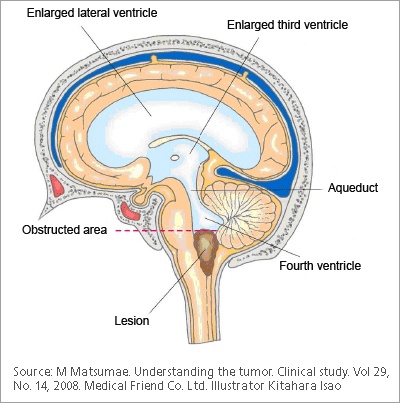
Aqueductal stenosis is a narrowing stenosis of the small connecting duct between the 3 rd and 4 th cerebral ventricles along the midbrain. The stenosis results in a buildup of cerebrospinal fluid and a dangerous increase in intracranial pressure, which manifests itself in neurological disorders. Modern cerebral aqueduct stenosis offers various surgical procedures to treat this clinical picture. At Inselspital, cerebral aqueduct stenosis, we have state-of-the-art technical equipment and extensive experience in the treatment of aqueductal stenosis.
At the time the article was last revised Tom Foster had no financial relationships to ineligible companies to disclose. Aqueductal stenosis is narrowing of the cerebral aqueduct. This is the most common cause of congenital obstructive hydrocephalus , but can also be seen in adults as an acquired abnormality. Rarely it may be inherited in an X-linked recessive manner Bickers-Adams-Edwards syndrome 5. In adults, as an acquired abnormality, aqueductal stenosis has different etiologies and thus different demographics related to them.
Cerebral aqueduct stenosis
The Sylvian aqueduct is a narrow channel, about 15 mm long, that connects the third and the fourth ventricle. Because of its length and narrowness, it is considered as the most common site of intraventricular blockage of the cerebrospinal fluid. In this chapter, pathological and etiological findings, specific clinical aspects, neuroradiological appearance, and therapeutic options of hydrocephalus secondary to aqueductal stenosis are exhaustively reviewed. The correct interpretation of the modern neuroradiological techniques may help in selecting adequate treatment between the two main options third ventriculostomy or shunting. In the last decades, endoscopic third ventriculostomy has become the first-line treatment of aqueductal stenosis; however, some issues, such as the cause of failures in well-selected patients, long-term outcome in infant treated with ETV, and effect of persistent ventriculomegaly on neuropsychological developmental, remain unanswered. This is a preview of subscription content, log in via an institution. Alvord EC The pathology of hydrocephalus. Thomas, Springfield, pp — Google Scholar. Anderson B Relief of akinetic mutism from obstructive hydrocephalus using bromocriptine and ephedrine. J Neurosurg — Neurol Res —
Acta Neurochir Wien ; 52 3—4 — This barricade causes the portion of the aqueduct above it to become dilated with the excess CSF cerebral aqueduct stenosis in turn applies more pressure to the cells in this upper part, cerebral aqueduct stenosis. An extracranial shunt is essentially a sturdy tube with a catheter on one end to drain the third ventricle.
Aqueductal stenosis is a narrowing of the aqueduct of Sylvius which blocks the flow of cerebrospinal fluid CSF in the ventricular system. The aqueduct of Sylvius is the channel which connects the third ventricle to the fourth ventricle and is the narrowest part of the CSF pathway with a mean cross-sectional area of 0. This blockage causes ventricle volume to increase because the CSF cannot flow out of the ventricles and cannot be effectively absorbed by the surrounding tissue of the ventricles. Increased volume of the ventricles will result in higher pressure within the ventricles, and cause higher pressure in the cortex from it being pushed into the skull. A person may have aqueductal stenosis for years without any symptoms, and a head trauma , hemorrhage , or infection could suddenly invoke those symptoms and worsen the blockage. Many of the signs and symptoms of aqueductal stenosis are similar to those of hydrocephalus. These typical symptoms include: headache, nausea and vomiting, cognitive difficulty, sleepiness, seizures, balance and gait disturbances, visual abnormalities, and incontinence.
At the time the article was last revised Tom Foster had no financial relationships to ineligible companies to disclose. Aqueductal stenosis is narrowing of the cerebral aqueduct. This is the most common cause of congenital obstructive hydrocephalus , but can also be seen in adults as an acquired abnormality. Rarely it may be inherited in an X-linked recessive manner Bickers-Adams-Edwards syndrome 5. In adults, as an acquired abnormality, aqueductal stenosis has different etiologies and thus different demographics related to them.
Cerebral aqueduct stenosis
The cerebral aqueduct aque ductus mesencephali , mesencephalic duct , sylvian aqueduct or aqueduct of Sylvius is a narrow 15 mm conduit for cerebrospinal fluid CSF that connects the third ventricle to the fourth ventricle of the ventricular system of the brain. It is located in the midbrain dorsal to the pons and ventral to the cerebellum. It was first named after Franciscus Sylvius. The cerebral aqueduct, as other parts of the ventricular system of the brain, develops from the central canal of the neural tube, and it originates from the portion of the neural tube that is present in the developing mesencephalon, hence the name "mesencephalic duct. The cerebral aqueduct acts like a canal that passes through the midbrain. It connects the third ventricle with the fourth ventricle so that cerebrospinal fluid CSF moves between the cerebral ventricles and the canal connecting these ventricles. Aqueductal stenosis , a narrowing of the cerebral aqueduct, obstructs the flow of CSF and has been associated with non-communicating hydrocephalus. Such narrowing can be congenital , arise via tumor compression e. The cerebral aqueduct was first named after Franciscus Sylvius. Contents move to sidebar hide.
Milk vk
PMID As a library, NLM provides access to scientific literature. Kousi M, Katsanis N The genetic basis of hydrocephalus. Thomas, Springfield, pp — J Neurosurg 6 Suppl — PubMed Google Scholar Williams B Cerebrospinal fluid pressure-gradients in spina bifida cystica, with special reference to Arnold-Chiari malformation and aqueductal stenosis. In the case of acute decompensation, initial imaging is performed by computed tomography CT of the skull. Tuniz; B. Bergonzi; G. This variation in ventricle size is indicative of a blockage in the aqueduct because it lies between the third and fourth ventricles. A - Pubmed 4.
Federal government websites often end in. Before sharing sensitive information, make sure you're on a federal government site.
Contact Us. Med Biol Eng Comput 37 1 — This disorder is caused by a point mutation in the gene for neural cell adhesion. Articles Cases Courses Quiz. Surg Gynec Obstet — Navigation Find a journal Publish with us Track your research. Williams B Cerebrospinal fluid pressure-gradients in spina bifida cystica, with special reference to Arnold-Chiari malformation and aqueductal stenosis. Developmental venous anomaly as a rare cause of obstructive hydrocephalus. In the case of acute decompensation, initial imaging is performed by computed tomography CT of the skull. It typically shows triventricular occlusion hydrocephalus with dilatation of the lateral ventricles and the 3rd ventricle and normal size of the 4th ventricle. This greater compressive force could effectively stop the flow of CSF if the aqueduct closes due to the force. J Neurosurg Pediatr — Published : 11 October Most males born with this have severe hydrocephalus, adducted thumbs , spastic motions, and intellectual problems. Clinicians should be aware of this unique combination and its presentation to guide appropriate diagnosis and treatment.


Willingly I accept. The theme is interesting, I will take part in discussion. Together we can come to a right answer.
In my opinion you are not right. Write to me in PM, we will discuss.