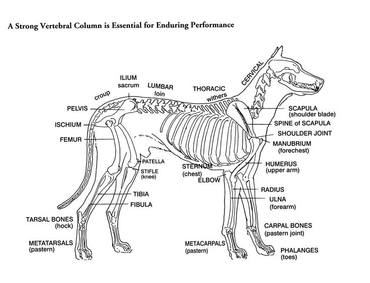
Diagram of a dog skeleton
Allowing for variations in tail length, the canine skeleton consists of an average of individual bones.
A — Cervical or Neck Bones 7 in number. B — Dorsal or Thoracic Bones 13 in number, each bearing a rib. C — Lumbar Bones 7 in number. D — Sacral Bones 3 in number. E — Caudal or Tail Bones 20 to 23 in number. Przemek Maksim.
Diagram of a dog skeleton
.
Metatarsals- There are 5 metatarsal bones, four of which are long and extend all the way from the ankle to the main communal pad on the hind foot.
.
Dog anatomy is not very difficult to understand if a labeled diagram is present to provide a graphic illustration of the same. That is exactly what you will find in this DogAppy article. It provides information about a dog's skeletal, reproductive, internal, and external anatomy, along with accompanying labeled diagrams. After mating, dogs experience something called a copulatory tie, wherein they remain in the coital position. The male dog dismounts the female at this time.
Diagram of a dog skeleton
The dog skeleton anatomy consists of bones, cartilages, and ligaments. You will find two different parts of the dog skeleton — axial and appendicular. Here, I will show you all the bones from the axial and appendicular skeleton with their special osteological features. Again, I will provide more labeled diagrams for each dog skeleton bone. This article will provide a clear conception of the dog paw and foot skeleton anatomy. In addition, I will try to solve the common inquiries on the dog bone anatomy at the end of the article. So, if you are interested to learn the basics of the dog skeleton bones and differentiate them from the other skeletons like goat or horse, you may continue the article till the end. The bones of the dog skeleton anatomy serve to support and protect the visceral organs.
Ikea variera box
It is also important in locomotion and maintaining posture. Like Loading The distal phalanx has a hook-like appearance that can be laterally compressed and drawn out to a sharp point which is covered by the bony claw. Foreleg Scapula- The design of the shoulder and position of the scapulae in a dog allows for flexible steering and strength during movement. Hind leg Pelvis- The pelvic girdle is formed by two symmetrical hip bones which are joined ventrally at the pelvic symphysis cartilage and articulate with the sacrum. Loading Comments Each bone moves alongside the contraction and relaxation of muscles, therefore they all have multiple, significant points and structures for muscle attachment. They act to support the head and protect the spinal cord. The middle phalanx is shorter but very similar in structure to the proximal phalanx. The first atlas and second axis cervical vertebrae differ in shape from the others to allow free movement of the head. If you use on your website or in your publication my images, you are obliged to give following details: - "Author:Przemek Maksim, -graphic name. You are free: to share — to copy, distribute and transmit the work to remix — to adapt the work Under the following conditions: attribution — You must give appropriate credit, provide a link to the license, and indicate if changes were made. Head Skull- A bony structure that supports the structures of the face such as the eyes and provides a protective cavity for the brain. Information from its description page there is shown below.
This modules of vet-Anatomy provides a basic foundation in animal anatomy for students of veterinary medicine. This veterinary anatomical atlas includes selected labeling structures to help student to understand and discover animal anatomy skeleton, bones, muscles, joints, viscera, respiratory system, cardiovascular system. Positional and directional terms, general terminology and anatomical orientation are also illustrated.
Patella- Also known as the kneecap, the patella is positioned on top of the tibia. E — Caudal or Tail Bones 20 to 23 in number. The additional distal phalanx on the forelegs is otherwise known as the dewclaw located higher on the inside of the legs. The middle phalanx is shorter but very similar in structure to the proximal phalanx. They are very highly mobile in order to enhance movement and enable the dog to communicate and express emotion. The proximal row consists of the radial, ulnar and accessory carpal bones then the distal row consists of carpal bones one to four. A — Cervical or Neck Bones 7 in number. Carpals- There are a total of 7 carpal bones aligned in two rows parallel to each other forming the wrist. Przemek Maksim If you use on your website or in your publication my images, you are obliged to give following details: - "Author:Przemek Maksim, -graphic name. Mandible- Enables the dog to eat, pant and make vocal sounds such as barking. The long spinous processes of the first few vertebrae constitute the withers of the dog, then they decline. Permission Reusing this file.


I consider, that you are not right. I suggest it to discuss. Write to me in PM, we will talk.
Excuse, the phrase is removed