Diffusion tensor imaging
Federal government websites often end in.
Diffusion Tensor Imaging DTI studies are increasingly popular among clinicians and researchers as they provide unique insights into brain network connectivity. However, in order to optimize the use of DTI, several technical and methodological aspects must be factored in. These include decisions on: acquisition protocol, artifact handling, data quality control, reconstruction algorithm, and visualization approaches, and quantitative analysis methodology. Herein, we provide a straightforward hitchhiker's guide, covering all of the workflow's major stages. Ultimately, this guide will help newcomers navigate the most critical roadblocks in the analysis and further encourage the use of DTI.
Diffusion tensor imaging
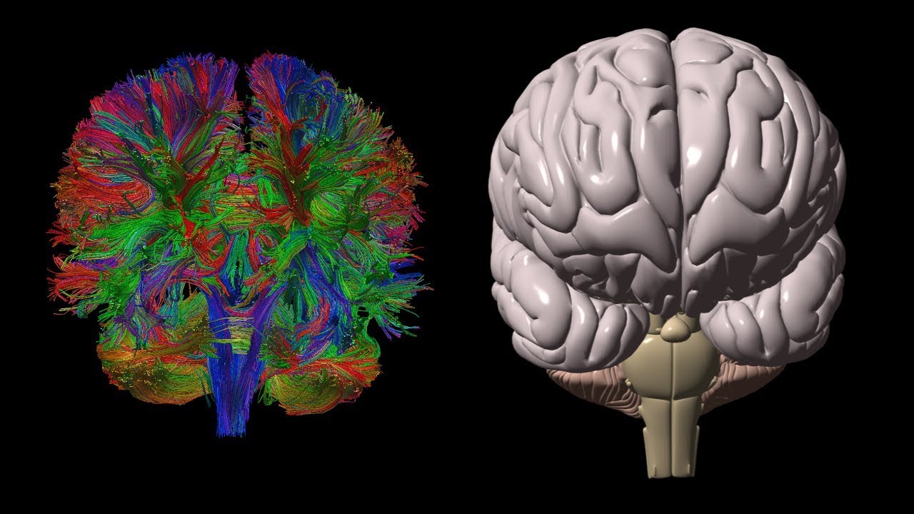
At the time the article was last revised Rohit Sharma had no financial relationships to ineligible companies to disclose. Diffusion tensor imaging DTI is an MRI technique that uses anisotropic diffusion to estimate the axonal white matter organization of the brain. Fiber tractography FT is a 3D reconstruction technique to assess neural tracts using data collected by diffusion tensor imaging. Diffusion-weighted imaging DWI is based on the measurement of thermal Brownian motion of water molecules. Within cerebral white matter, water molecules tend to diffuse more freely along the direction of axonal fascicles rather than across them. Such directional dependence of diffusivity is termed anisotropy. This direction of maximum diffusivity along the white-matter fibers is projected in the final image. However, its use for the assessment of highly-organized body systems outside the CNS, where anisotropy can facilitate early detection of pathology, has been gaining favor in recent years. Alzheimer disease - detection of early disease. FA reflects the directionality of molecular displacement by diffusion and varies between 0 isotropic diffusion and 1 infinite anisotropic diffusion. FA value of CSF is 0. MD reflects the average magnitude of molecular displacement by diffusion.
C 10, L55—L In: Weickert Joachim, Hagen Hans.
Diffusion tensor imaging DTI allows a live look into the microstructure of white matter in the brain and is an important complement to volumetric studies of specific structures such as the amygdala. DTI may be particularly informative for the study of autism because it has been speculated that white matter the connections between neurons defects may be even more pronounced than gray matter defects for affected individuals. Furthermore, in contrast to gray matter, white matter volume continues to increase across childhood and adolescence, thus allowing for analyses of growth curves and changes specific to the microstructure of axons. See papers by Courchesne, et al. Studies that utilize DTI technology generally describe two characteristics of white matter within a particular "voxel" in the brain:.
Diffusion MRI is used widely to probe microstructural alterations in neurological and psychiatric disease. However, ageing and neurodegeneration are also associated with atrophy, which leads to artefacts through partial volume effects due to cerebrospinal-fluid contamination CSFC. The aim of this study was to explore the influence of CSFC on apparent microstructural changes in mild cognitive impairment MCI at several spatial levels: individually reconstructed tracts; at the level of a whole white matter skeleton tract-based spatial statistics ; and histograms derived from all white matter. We corrected for CSFC using a post-acquisition voxel-by-voxel approach of free-water elimination. Tracts varied in their susceptibility to CSFC. The apparent pattern of tract involvement in disease shifted when correction was applied. Both spurious group differences, driven by CSFC, and masking of true differences were observed.
Diffusion tensor imaging
The precise characterization of cerebral thrombi prior to an interventional procedure can ease the procedure and increase its success. This study investigates how well cerebral thrombi can be characterized by computed tomography CT , magnetic resonance MR and histology, and how parameters obtained by these methods correlate with each other as well as with the interventional procedure and clinical parameters. Cerebral thrombi of 25 patients diagnosed by CT with acute ischemic stroke were acquired by mechanical thrombectomy and, subsequently, scanned by a high spatial-resolution 3D MRI including T 1 -weighted imaging, apparent diffusion coefficient ADC , T 2 mapping and then finally analyzed by histology.
Aldis joplin mo
FA value of CSF is 0. Figure 1. OCLC Geometrically constrained two-tensor model for crossing tracts in DWI. Human acute cerebral ischemia: detection of changes in water diffusion anisotropy by using MR imaging. A unified framework for clustering and quantitative analysis of white matter fiber tracts. Within cerebral white matter, water molecules tend to diffuse more freely along the direction of axonal fascicles rather than across them. Behrens, T. First, an anatomic scan is performed. Acute stroke Pregnancy.
Diffusion-weighted magnetic resonance imaging DWI or DW-MRI is the use of specific MRI sequences as well as software that generates images from the resulting data that uses the diffusion of water molecules to generate contrast in MR images.
Putting the two together, we get the diffusion equation :. The imaging of this property is an extension of diffusion MRI. A comparative study of acquisition schemes for diffusion tensor imaging using MRI. Intraoperative visualization of the pyramidal tract by diffusion-tensor-imaging-based fiber tracking. Clinically, trace-weighted images have proven to be very useful to diagnose vascular strokes in the brain, by early detection within a couple of minutes of the hypoxic edema. In particular, FA is highly sensitive to microstructural changes, but not very specific to the type of changes e. Differentiation of recurrent brain tumor versus radiation injury using diffusion tensor imaging in patients with new contrast-enhancing lesions. It is also used more and more in the staging of non-small-cell lung cancer , where it is a serious candidate to replace positron emission tomography as the 'gold standard' for this type of disease. Imaging 29, — Typical DTI workflow. In fact, this sensitivity, providing diffusion summary measures and tissue fiber orientation, has made DTI widely used as a clinical tool, especially in conditions where abnormalities in WM are expected Sundgren et al. Pfefferbaum, A. In fact, the high dimensionality of the data and the complex associations in diffusion tensors domain make this step quite problematic.


0 thoughts on “Diffusion tensor imaging”