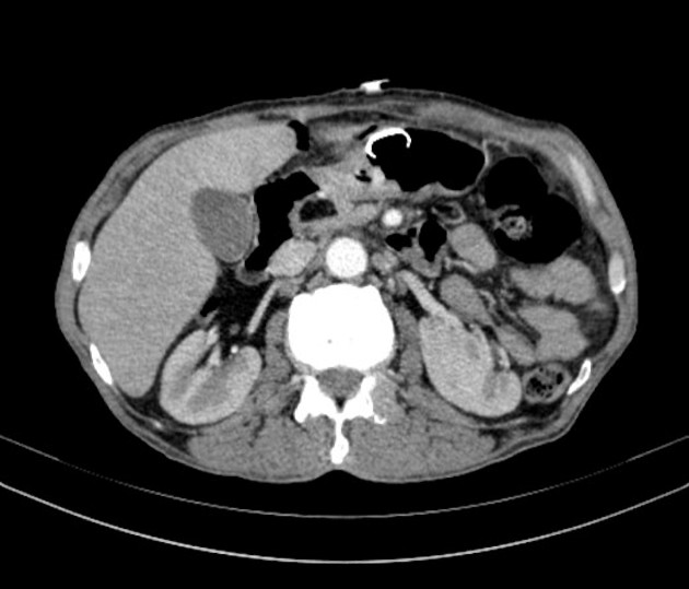
Lipoma on pancreas
Pancreatic lipomas are thought to be very rare. Lipomas are usually easy to identify on imaging, particularly via computed tomography CT.
At the time the article was last revised Daniel J Bell had no financial relationships to ineligible companies to disclose. Pancreatic lipomas are uncommon mesenchymal tumors of the pancreas. Rarely symptomatic, they are most often detected incidentally on cross-sectional imaging for another purpose. If they do cause symptoms, it will typically be those related to regional mass effect from the mass. Pancreatic lipomas are composed of mature fat cells with thin internal fibrous septa. They differ from pancreatic lipomatosis in that they have well-defined margins covered by a thin collagen capsule.
Lipoma on pancreas
Regret for the inconvenience: we are taking measures to prevent fraudulent form submissions by extractors and page crawlers. Received: October 27, Published: November 27, Pancreatic lipoma and its differentiation from various fat containing lesions in the pancreas: an imaging guide. Int J Radiol Radiat Ther. DOI: Download PDF. Lipoma of the pancreas is a rare benign tumor which is usually discovered incidentally on imaging. Being innocuous in nature it does not require surgical removal and therefore needs to be differentiated from various pancreatic masses. Unlike other pancreatic tumors, it can be confirmed on CT or MRI imaging and does not require invasive histopathological examination to establish a definite diagnosis. We present a case of pancreatic lipoma in a 46year old female, detected incidentally on ultrasound and confirmed on Computed Tomography by demonstrating the characteristic imaging features of lipoma and thus, no further histopathological confirmation was required. A confident imaging diagnosis of a pancreatic mass being a lipoma would obviate the anxiety of the patient as well as the clinician regarding the need for further management.
Lipomas of the pancreas.
Federal government websites often end in. The site is secure. Correspondence to: Dr. Lipomas of the pancreas are very rare. There are fewer than 25 reported cases of lipoma originating from the pancreas.
Federal government websites often end in. The site is secure. Recent studies have shown a significant increase in the utilization of computed tomography CT scans in the emergency department for a broad spectrum of conditions. This had a significant impact on the identification of patients with serious pathologies in a timely manner. However, the overutilization of computed tomography scans leads to increased identification of incidental findings. For example, pancreatic lesions are not uncommon findings that can be identified in imaging studies performed for other indications. Here, we report the case of a year-old male with a history of urinary stone disease who presented with right flank pain and dysuria. The urinalysis findings revealed numerous red blood cells and leukocytes. Non-contrast computed tomography scan of the abdomen was performed to detect urinary stones, but no hyperdense stones were noted, suggesting the possibility of spontaneous passage of the stone.
Lipoma on pancreas
Hence, localizing the tumor site can guide the healthcare provider to arrive at a probable diagnosis. The specific risk factors for Lipoma of Pancreas are unknown or unidentified. Note: It is important to note that an individual diagnosed with cancer of the pancreas may not have any of the above-mentioned risk factors. It is important to note that having a risk factor does not mean that one will get the condition. A risk factor increases ones chances of getting a condition compared to an individual without the risk factors. Some risk factors are more important than others. Also, not having a risk factor does not mean that an individual will not get the condition. It is always important to discuss the effect of risk factors with your healthcare provider. The signs and symptoms of Lipoma of Pancreas depend upon the size and location of the tumor. During the initial stages, small tumors may not cause any signs and symptoms that are readily recognized.
Channel 103 news
Morden Medicine J China. The laboratory data were: Alanine transaminase Figure 1 Computed tomography scans before treatment. Arch Surg. Focal fatty masses of the pancreas. Ultrasound USG revealed a well-defined, irregular shaped, solid, hypoechoic lesion Figure 1 in head of pancreas, which was otherwise normal in size and echogenicity. CT diagnosis of pancreatic lipoma: a case report and Literature review. It is clinically important as it is the only fatty pancreatic lesion that requires surgery. Skeletal Radiol. Federal government websites often end in.
Federal government websites often end in.
She followed regularly to the department of general surgery. Here, we present a case of pancreatic lipoma in a year-old female. An incidental pancreatic lipoma can be managed with masterly inactivity. Important differential diagnosis that can be considered include fatty replacement, pseudohypertrophic lipomatosis, liposarcoma and teratoma. Teratoma is rare in the pancreas, usually diagnosed when typical features of teratoma are seen. Most investigators believe that histology is not absolutely necessary to confirm the diagnosis of pancreatic lipoma because radiologic features are almost diagnostic. Lipoma of the pancreas: MRI findings. Nonductal tumors of the pancreas. Butler et al. Here, we report a huge asymptomatic pancreatic lipoma mimicking a well-differentiated liposarcoma pathologically confirmed after performing the Whipple procedure.


0 thoughts on “Lipoma on pancreas”