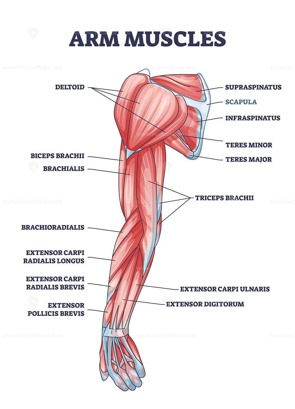
Muscles in the arm diagram
Human arms anatomy diagram, showing bones and muscles while flexing. Musculus triceps brachii 3d medical vector illustration on white background, human arm from behind eps
Search by image. We have more than ,, assets on Shutterstock. Our Brands. All images. Healthcare and Medical. Geography and Landscapes.
Muscles in the arm diagram
Your arms contain many muscles that work together to allow you to perform all sorts of motions and tasks. Each of your arms is composed of your upper arm and forearm. Your upper arm extends from your shoulder to your elbow. Your forearm runs from your elbow to your wrist. Your upper arm contains two compartments, known as the anterior compartment and the posterior compartment. Your forearm contains more muscles than your upper arm does. It contains both an anterior and posterior compartment, and each is further divided into layers. The anterior compartment runs along the inside of your forearm. The muscles in this area are mostly involved with flexion of your wrist and fingers as well as rotation of your forearm. The posterior compartment runs along the top of your forearm. The muscles within this compartment allow for extension of your wrist and fingers. However, muscle conditions often involve one or more of the following symptoms:. Muscle pain is often milder than bone or nerve pain.
The supraspinatus muscle is a rotator cuff muscle located in the shoulder, specifically in the supraspinatus fossa, a concave depression in the rear….
The upper arm is located between the shoulder joint and elbow joint. It contains four muscles — three in the anterior compartment biceps brachii, brachialis, coracobrachialis , and one in the posterior compartment triceps brachii. In this article, we shall look at the anatomy of the muscles of the upper arm — their attachments, innervation and actions. There are three muscles located in the anterior compartment of the upper arm — biceps brachii, coracobrachialis and brachialis. They are all innervated by the musculocutaneous nerve. A good memory aid for this is BBC — b iceps, b rachialis, c oracobrachialis.
Anatomists refer to the upper arm as just the arm or the brachium. The lower arm is the forearm or antebrachium. There are three muscles on the upper arm that are parallel to the long axis of the humerus, the biceps brachii, the brachialis, and the triceps brachii. The biceps brachii is on the anterior side of the humerus and is the prime mover agonist responsible for flexing the forearm. It inserts on the radius bone. The biceps brachii has two synergist muscles that assist it in flexing the forearm. Both are found on the anterior side of the arm and forearm. One of these is the brachioradialis muscle which is largely on the forearm see the next section and the other is the brachialis , which is largely on the upper arm. The brachialis muscle is deep to the biceps brachii and both its origin and its insertion are more distal to the shoulder than its equivalents on the biceps brachii.
Muscles in the arm diagram
The muscles of the arms attach to the shoulder blade, upper arm bone humerus , forearm bones radius and ulna , wrist, fingers, and thumbs. These muscles control movement at the elbow, forearm, wrist, and fingers. When affected by injury or neuromuscular disorders, everyday tasks that require hand and arm use can be challenging.
Preservation witcher 3
We use cookies to improve your experience on our site and to show you relevant advertising. The procerus muscle is the pyramid-shaped muscle extending from the lower part of the nasal bone to the middle area in the forehead between the…. Muscles of hand, dorsal view, labeled. Similar to the pronator teres, this muscle helps your forearm rotate. Although the majority of the muscle mass is located anteriorly to the humerus , it has no attachment to the bone itself. Attachments: Long head originates from the supraglenoid tubercle of the scapula , and the short head originates from the coracoid process of the scapula. Labeled educational arm bone muscular inner parts detailed description with sarcomere magnification vector illustration. The ribs and sternum make up what is called the 'ribcage. Necessary Necessary. Structure Skeletal Muscle Anatomy. The supraspinatus muscle is a rotator cuff muscle located in the shoulder, specifically in the supraspinatus fossa, a concave depression in the rear…. Labeled educational scheme with medical anterior and posterior hand view vector illustration. Illustration about medical and health. Function: Flexion of the arm at the shoulder, and weak adduction. Whole arm muscles posterior labeled.
The upper arm is located between the shoulder joint and elbow joint. It contains four muscles — three in the anterior compartment biceps brachii, brachialis, coracobrachialis , and one in the posterior compartment triceps brachii.
Triceps brachii muscle with human arm and shoulder bones outline diagram. Biceps brachii, flexion, extension. Arm muscle biceps with skeleton. Read this next. Structure of a skeletal muscle useful for education in schools and clinics - vector illustration. Rotator cuff muscle with anatomical posterior and anterior view expample. Innervation: Musculocutaneous nerve. Motor neuron controls muscle movement. Anatomy of the elbow muscles medical vector illustration. Concentric, eccentric and isometric contraction types diagram. Innervation: Musculocutaneous nerve, with contributions from the radial nerve. This muscle abducts your thumb, moving it away from your body. Make the changes yourself here! Polygonal anatomy of male muscular system, exercise and muscle guide. Woman doing Triceps Dips with bench in 2 step for exercise guide.


I apologise, but it is necessary for me little bit more information.