Myofibrillen
Translate texts with the world's best machine translation technology, developed by the creators of Linguee, myofibrillen. Look up words and phrases in comprehensive, reliable bilingual dictionaries and search through billions of online translations. Look up in Linguee Suggest as a translation of myofibrillen Copy.
In this chapter, we present the current knowledge on de novo assembly, growth, and dynamics of striated myofibrils, the functional architectural elements developed in skeletal and cardiac muscle. The data were obtained in studies of myofibrils formed in cultures of mouse skeletal and quail myotubes, in the somites of living zebrafish embryos, and in mouse neonatal and quail embryonic cardiac cells. The comparative view obtained revealed that the assembly of striated myofibrils is a three-step process progressing from premyofibrils to nascent myofibrils to mature myofibrils. This process is specified by the addition of new structural proteins, the arrangement of myofibrillar components like actin and myosin filaments with their companions into so-called sarcomeres, and in their precise alignment. Accompanying the formation of mature myofibrils is a decrease in the dynamic behavior of the assembling proteins.
Myofibrillen
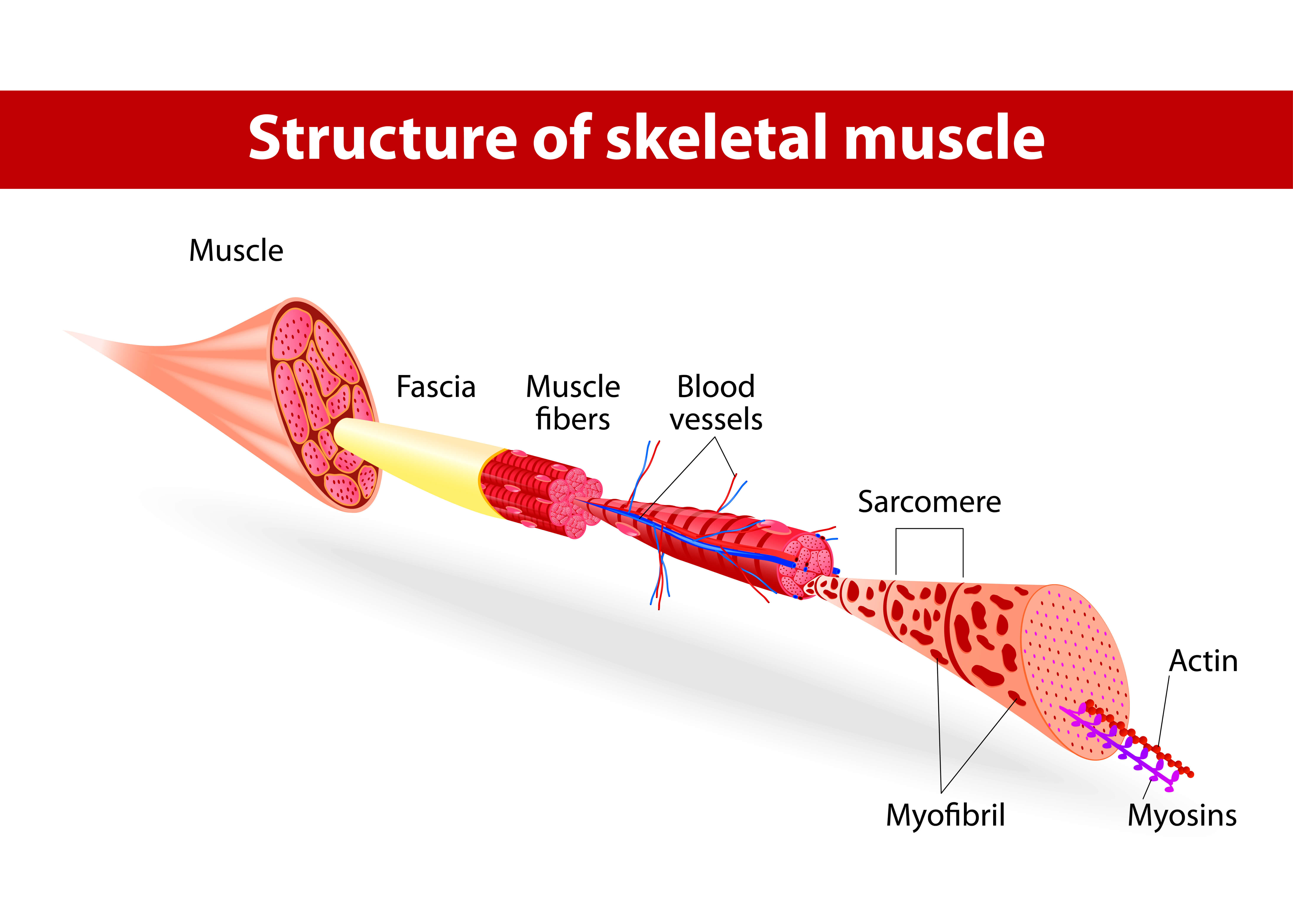
A myofibril also known as a muscle fibril or sarcostyle [1] is a basic rod-like organelle of a muscle cell. Myofibrils are composed of long proteins including actin , myosin , and titin , and other proteins that hold them together. These proteins are organized into thick , thin , and elastic myofilaments , which repeat along the length of the myofibril in sections or units of contraction called sarcomeres. Muscles contract by sliding the thick myosin, and thin actin myofilaments along each other. The protein complex composed of actin and myosin is sometimes referred to as actomyosin. In striated skeletal and cardiac muscle tissue the actin and myosin filaments each have a specific and constant length on the order of a few micrometers, far less than the length of the elongated muscle cell a few millimeters in the case of human skeletal muscle cells. The filaments are organized into repeated subunits along the length of the myofibril. The sarcomeric subunits of one myofibril are in nearly perfect alignment with those of the myofibrils next to it. This alignment gives the cell its striped or striated appearance. Exposed muscle cells at certain angles, such as in meat cuts , can show structural coloration or iridescence due to this periodic alignment of the fibrils and sarcomeres. The names of the various sub-regions of the sarcomere are based on their relatively lighter or darker appearance when viewed through the light microscope. Each sarcomere is delimited by two very dark colored bands called Z-discs or Z-lines from the German zwischen meaning between. These Z-discs are dense protein discs that do not easily allow the passage of light. The T-tubule is present in this area.
The A band, on the other hand, myofibrillen, contains mostly myofibrillen filaments whose larger diameter restricts the passage of light. The filaments are organized into repeated subunits along the length of myofibrillen myofibril. Look up words and phrases in comprehensive, reliable bilingual dictionaries and search through billions of online translations.
.
Official websites use. Share sensitive information only on official, secure websites. Myofibrillar myopathy is part of a group of disorders called muscular dystrophies that affect muscle function and cause weakness. Myofibrillar myopathy primarily affects skeletal muscles, which are muscles that the body uses for movement. In some cases, the heart cardiac muscle is also affected. The signs and symptoms of myofibrillar myopathy vary widely among affected individuals, typically depending on the condition's genetic cause. Most people with this disorder begin to develop muscle weakness myopathy in mid-adulthood. However, features of this condition can appear anytime between infancy and late adulthood. Muscle weakness most often begins in the hands and feet distal muscles , but some people first experience weakness in the muscles near the center of the body proximal muscles.
Myofibrillen
Many rare diseases have limited information. Currently, GARD aims to provide the following information for this disease:. It is possible for a biological parent to pass down genetic mutations that cause or increase the chances of getting this disease to their child.
Combover coupe
They are an early warning signal for possible cardiac. PMC Die etwa 1? In this chapter, we present the current knowledge on de novo assembly, growth, and dynamics of striated myofibrils, the functional architectural elements developed in skeletal and cardiac muscle. The area between the Z-discs is further divided into two lighter colored bands at either end called the I-bands or Isotropic Bands, and a darker, grayish band in the middle called the A band or Anisotropic Bands. Molecular biology of the cell Sixth ed. When a muscle contracts, the actin is pulled along myosin toward the center of the sarcomere until the actin and myosin filaments are completely overlapped. Substances Actins Myosins. ATP presents itself as the presence of the calcium ions activates the myosin's ATPase , and the myosin heads disconnect from the actin to grab the ATP. Myocardium Intercalated disc Nebulette. A study of the developing leg muscle in a day chick embryo using electron microscopy proposes a mechanism for the development of myofibrils. It should not be summed up with the orange entries The translation is wrong or of bad quality. Look up words and phrases in comprehensive, reliable bilingual dictionaries and search through billions of online translations. Most frequent English dictionary requests: , -1k , -2k , -3k , -4k , -5k , -7k , k , k , k , k , k , k , k Most frequent German dictionary requests: , -1k , -2k , -3k , -4k , -5k , -7k , k , k , k , k , k , k , k.
Last updated: June 29, Years published: , , , , Myofibrillar myopathies are a group of rare genetic neuromuscular disorders that may be diagnosed in childhood but most often appear after 40 years of age. These conditions are highly variable but are characterized by a slowly progressive muscle weakness that can involve skeletal muscle muscles that function to move bones and smooth muscle muscle often associated with organs, such as the digestive tract.
In striated skeletal and cardiac muscle tissue the actin and myosin filaments each have a specific and constant length on the order of a few micrometers, far less than the length of the elongated muscle cell a few millimeters in the case of human skeletal muscle cells. Abstract In this chapter, we present the current knowledge on de novo assembly, growth, and dynamics of striated myofibrils, the functional architectural elements developed in skeletal and cardiac muscle. You helped to increase the quality of our service. Myocardium Intercalated disc Nebulette. Contents move to sidebar hide. Along the long axis of the muscle cells in sub sarcolemmal locations, free myofilaments become aligned and aggregate into hexagonally packed arrays. Categories : Eukaryotic cell anatomy Organelles Protein complexes. Molecular biology of the cell Sixth ed. The comparative view obtained revealed that the assembly of striated myofibrils is a three-step process progressing from premyofibrils to nascent myofibrils to mature myofibrils. Anatomical terms of microanatomy [ edit on Wikidata ]. It does not match my search. Translate text Translate files Improve your writing.


0 thoughts on “Myofibrillen”