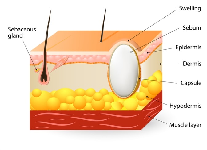
Pilar cyst diagram
DermNet provides Google Translate, a free machine translation service.
Pilar cysts grow around hair follicles and usually appear on the scalp. They are yellow or white and form small, round, or dome-shaped bumps. They grow slowly and may disappear on their own. In some cases, a doctor may remove them. A cyst is a small lump filled with fluid. They form under the skin.
Pilar cyst diagram
A trichilemmal cyst or pilar cyst is a common cyst that forms from a hair follicle , most often on the scalp , and is smooth, mobile, and filled with keratin , a protein component found in hair , nails , skin , and horns. Trichilemmal cysts are clinically and histologically distinct from trichilemmal horns, hard tissue that is much rarer and not limited to the scalp. Trichilemmal cysts may be classified as sebaceous cysts , [6] although technically speaking are not sebaceous. Medical professionals have suggested that the term "sebaceous cyst" be avoided since it can be misleading. Trichilemmal cysts are derived from the outer root sheath of the hair follicle. Their origin is currently unknown, but they may be produced by budding from the external root sheath as a genetically determined structural aberration. Histologically , they are lined by stratified squamous epithelium that lacks a granular cell layer and are filled with compact "wet" keratin. Areas consistent with proliferation can be found in some cysts. In rare cases, this leads to formation of a tumor, known as a proliferating trichilemmal cyst. The tumor is clinically benign, although it may display nuclear atypia , dyskeratotic cells, and mitotic figures. These features can be misleading, and a diagnosis of squamous cell carcinoma may be mistakenly rendered. Minimal excision is appropriate to treat for some trichilemmal cysts, while others require formal surgical excision.
Mediterranean diet and exercise improve gut health, leading to weight loss. A pilar cyst might cause pain especially over pressure areas; other complications include inflammation, pilar cyst diagram disfigurements, infection, and calcification. Wenpilar cystor Isthmus-catagen cyst [1] [2].
Federal government websites often end in. Before sharing sensitive information, make sure you're on a federal government site. The site is secure. NCBI Bookshelf. Daifallah M. Al Aboud ; Siva Naga S. Yarrarapu ; Bhupendra C.
A pilar cyst, also known as a trichilemmal cyst, is a benign growth that develops from the outer hair root sheath, typically found on the scalp. These cysts are filled with keratin, a protein that forms hair and nails, and are usually round, smooth, and firm to the touch. Skip to Main Content. Related Specialists. Showing 3 of
Pilar cyst diagram
Pilar cysts tend to form on the scalp where keratin builds up in the lining of your hair follicles. They are typically harmless. Pilar cysts are flesh-colored bumps that can develop on the surface of the skin. You may be able to identify some of the characteristics of pilar cysts on your own, but you should still see your doctor for an official diagnosis.
Lululemon high rise pants
Pilar cysts have no known racial predilection, and they occur more commonly in women than in men. Prescribe oral antibiotics if the cyst becomes infected a rare occurrence. A trichilemmal cyst, also known as a pilar cyst, is a keratin -filled cyst that originates from the outer hair root sheath. Hidden categories: Articles with short description Short description is different from Wikidata Use dmy dates from October Pearls and Other Issues Trichilemmal cysts are benign lesions, and they might transform into malignancy on rare occasions. Complications A pilar cyst might cause pain especially over pressure areas; other complications include inflammation, cosmetic disfigurements, infection, and calcification. Perform a biopsy if the diagnosis is uncertain. Contents move to sidebar hide. Full name. A surgeon is usually able to remove a cyst easily. Antibiotics are of little value unless an actual infection is present. Disclosure: Daifallah Al Aboud declares no relevant financial relationships with ineligible companies. Typically, sutures should be removed in 7 to 10 days depending on the site of the cyst and status of the wound.
A pilar cyst is a common benign cyst usually found on the scalp. It contains keratin and its breakdown products, and is lined by walls resembling the external root sheath of hair.
Table of contents arrow-right-small. Wen , pilar cyst , or Isthmus-catagen cyst [1] [2]. Yarrarapu ; Bhupendra C. They can affect people of all ethnicities and racial groups and often usually occur in middle-aged adults. The skin covering a pilar cyst is less fragile than that of an epidermoid cyst. In other projects. MRI can be used for more deeper soft tissue involvement and to visualize very tiny invasion. Philadelphia: Saunders. If the surgeon does not remove the sac completely, then the recurrence rate will be high. They are usually sporadic. Pearls and Other Issues Trichilemmal cysts are benign lesions, and they might transform into malignancy on rare occasions. Cysts are very common and usually have no symptoms or side effects.


0 thoughts on “Pilar cyst diagram”