Sliding filament theory coloring answers
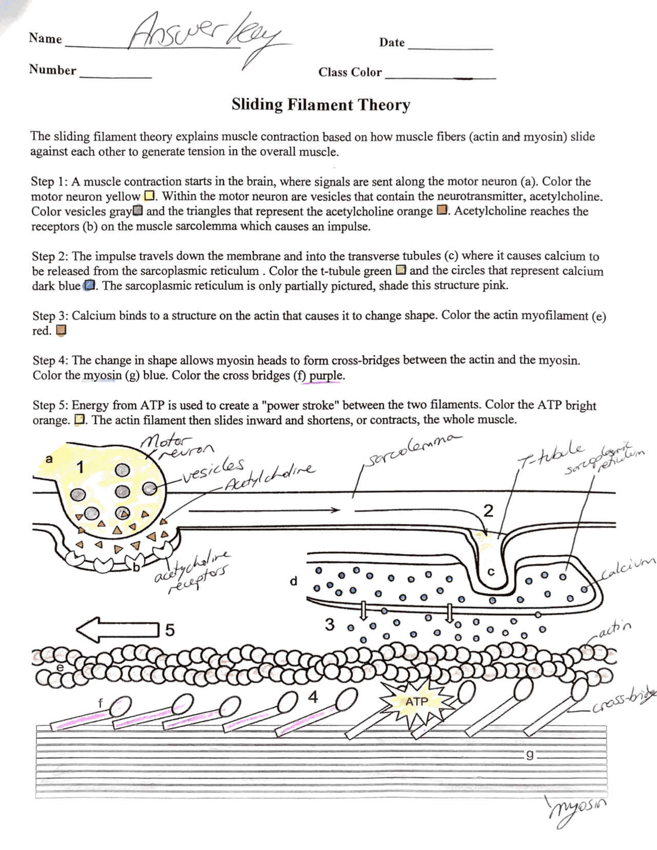
The sliding filament theory explains muscle contraction based on how muscle fibers actin and myosin slide against each other to generate tension in the overall muscle. Step 1 : A muscle contraction starts in the brain, where a signal is sent to the motor neuron a.
Sliding Filament Graphic - shows how actin and myosin interact, labeled in stages. Includes questions to answer. Sliding Filament Model slides - shows the steps of mucles contraction, students label a worksheet. Label Musscle Structure - includes entire muscle epimysium, perimysium, endomysium , then has a sarcomere labeling. Presented as Google slides, that students can drag labels to structures. Label Neuromuscular Junction - Google slide labeling that includes how a neuron interacts with a muscle motor unit. Sarcomere Coloring - black and white image shows the sarcomere and the actin and myosin filaments.
Sliding filament theory coloring answers
Each skeletal muscle is an organ that consists of various integrated tissues. These tissues include the skeletal muscle fibers, blood vessels, nerve fibers, and connective tissue. Each skeletal muscle has three layers of connective tissue called mysia that enclose it, provide structure to the muscle, and compartmentalize the muscle fibers within the muscle Figure Each muscle is wrapped in a sheath of dense, irregular connective tissue called the epimysium , which allows a muscle to contract and move powerfully while maintaining its structural integrity. The epimysium also separates muscle from other tissues and organs in the area, allowing the muscle to move independently. Inside each skeletal muscle, muscle fibers are organized into bundles, called fascicles , surrounded by a middle layer of connective tissue called the perimysium. This fascicular organization is common in muscles of the limbs; it allows the nervous system to trigger a specific movement of a muscle by activating a subset of muscle fibers within a fascicle of the muscle. Inside each fascicle, each muscle fiber is encased in a thin connective tissue layer of collagen and reticular fibers called the endomysium. The endomysium surrounds the extracellular matrix of the cells and plays a role in transferring force produced by the muscle fibers to the tendons. In skeletal muscles that work with tendons to pull on bones, the collagen in the three connective tissue layers intertwines with the collagen of a tendon. At the other end of the tendon, it fuses with the periosteum coating the bone. The tension created by contraction of the muscle fibers is then transferred though the connective tissue layers, to the tendon, and then to the periosteum to pull on the bone for movement of the skeleton. In other places, the mysia may fuse with a broad, tendon-like sheet called an aponeurosis , or to fascia, the connective tissue between skin and bones. Every skeletal muscle is also richly supplied by blood vessels for nourishment, oxygen delivery, and waste removal. In addition, every muscle fiber in a skeletal muscle is supplied by the axon branch of a somatic motor neuron, which signals the fiber to contract.
Energy from ATP is required for the myosin head to release from the actin filament—otherwise the myosin heads would remain in the same place, and the muscle would not contract. The impulse travels down membrane, or sarcolemma.
For complaints, use another form. Study lib. Upload document Create flashcards. Flashcards Collections. Documents Last activity. Add to Add to collection s Add to saved.
In the sliding filament model, the thick and thin filaments pass each other, shortening the sarcomere. Movement often requires the contraction of a skeletal muscle, as can be observed when the bicep muscle in the arm contracts, drawing the forearm up towards the trunk. The sliding filament model describes the process used by muscles to contract. It is a cycle of repetitive events that causes actin and myosin myofilaments to slide over each other, contracting the sarcomere and generating tension in the muscle. To understand the sliding filament model requires an understanding of sarcomere structure. A sarcomere is defined as the segment between two neighbouring, parallel Z-lines.
Sliding filament theory coloring answers
It starts with a signal from the nervous system. So it starts with a signal from your brain. The signal goes through your nervous system to your muscle. Your muscle contracts, and your bones move.
Why cant i download forza horizon 3
Step 3: Calcium binds to a structure on the actin that causes it to change shape. Your e-mail Input it if you want to receive answer. The actin filament then slides inward and shortens, or contracts, the whole muscle. The tension created by contraction of the muscle fibers is then transferred though the connective tissue layers, to the tendon, and then to the periosteum to pull on the bone for movement of the skeleton. Step 3: Calcium binds to the troponin-tropomyosin complex on the actin that causes it to change shape and move from the myosin binding site in the actin. Subjects Math. Also included in: Muscular System Unit Bundle. Step 5 : Energy from ATP is used to create a "power stroke" between the two filaments. Log In Join. Color the actin myofilament e red. Acetylcholine reaches the receptors b on the muscle sarcolemma which causes an impulse. Color the motor neuron a yellow.
Sliding Filament Graphic - shows how actin and myosin interact, labeled in stages.
What are the two filaments found in muscles? All 'Science'. Interactive Notebooks. Clip art. Bulletin board ideas. Service learning. Speech therapy. Interactive Link Questions Watch this video to learn more about macro- and microstructures of skeletal muscles. This download has two versions of the student worksheet 2 pages vs 1 pages and the answer key to the questions. What substance provides the energy for muscle contraction?


0 thoughts on “Sliding filament theory coloring answers”