Soft tissue density lesion meaning
Federal government websites often end in.
At the time the article was created Joachim Feger had no financial relationships to ineligible companies to disclose. At the time the article was last revised Daniel J Bell had no financial relationships to ineligible companies to disclose. Soft tissue masses or lesions are a common medical condition seen by primary care physicians, family physicians, surgeons and orthopedists. They include all outgrowths, both benign and malignant, arising from soft tissue Soft tissue masses are very common, with benign lesions being much more frequent than their malignant counterparts, outnumbering them by about to one Sarcomas can occur at any age and are generally more common in older people 1,2. However, the ratio of malignant versus benign soft tissue lesions is higher in children because benign lipomas and epidermal cysts are infrequent in that population 1.
Soft tissue density lesion meaning
Magnetic resonance imaging MRI is far superior to computed tomography CT for the visualization of soft tissue pathology because of greater soft tissue contrast and an overall improved tissue characterization based on signal behavior on different pulse sequences and relaxation parameters. Compared with MRI, CT is more sensitive for the diagnosis of both tiny soft tissue calcifications and air collections and facilitates differentiation between the two. For CT, the contrast characteristics of soft tissue disease depend on the relative proportions of fat, water, and mineral. Normal muscles are of soft tissue density and are separated from each other by fatty septa. In many muscle diseases, the muscle fibers become necrotic and degenerate or are replaced by fat and connective tissue. Fatty replacement of muscle may be complete and homogeneous or incomplete and inhomogeneous, but it is not characteristic for a specific disease. It is observed with muscular dystrophies, neuropathies, ischemias, and metabolic and systemic myopathies, as well as idiopathically Fig. CT is of little use in the differentiation of these conditions, but it may play an important role in the localization, distribution, and assessment of the extent of muscular involvement. CT is useful in the evaluation of soft tissue masses. It allows definition of the exact dimensions of a lesion and its relationship to nearby neurovascular structures and bone.
The pathologist uses laboratory technology to count the cells and determine their rate of growth. Fairly well to poorly defined soft tissue mass.
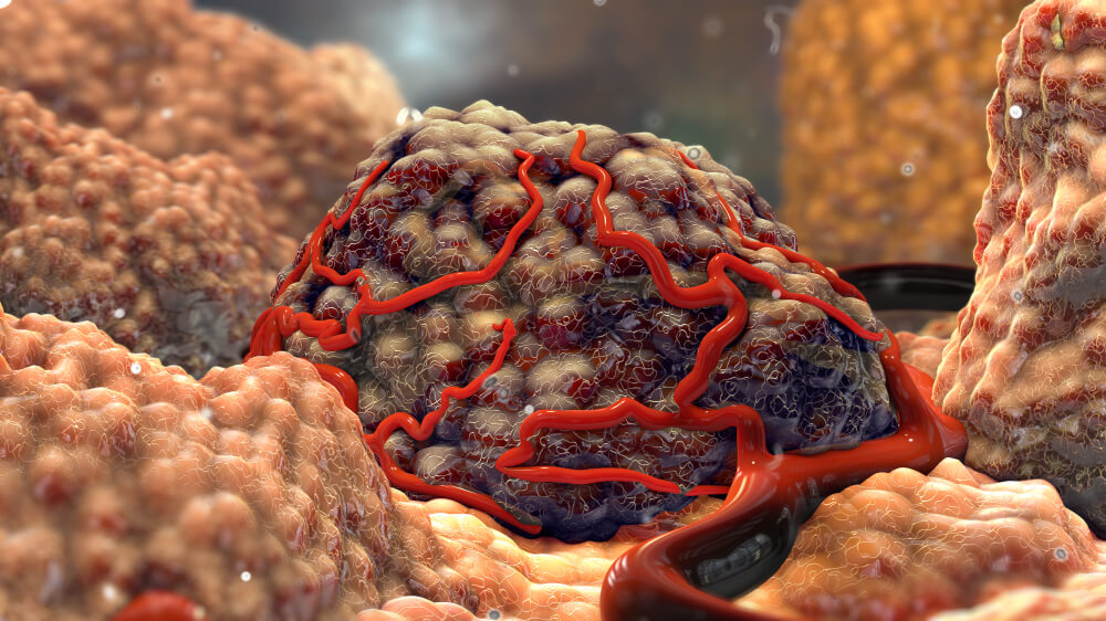
Soft tissue tumors, which are also called soft tissue masses, can be found anywhere in the body. Here are 10 important things you need to know about soft tissue tumors. You may be asking yourself, what is a soft tissue mass or tumor? A tumor in your soft tissue means that some fat, muscle, or other non-bone cells have multiplied in number more than they should have. Most of these begin in the fat cells.
Soft tissue lesions strike fear in many pathologists as they are uncommon and may be difficult to diagnose. Malignant soft tissue lesions, i. Sarcomas are malignancies derived from mesenchymal tissue. These include: [1]. These include: [2]. Most common: [3]. Components - overview: [7] [8]. Exceptions: [8].
Soft tissue density lesion meaning
A radiographic image is composed of a 'map' of X-rays that have either passed freely through the body or have been variably attenuated absorbed or scattered by anatomical structures. The denser the tissue, the more X-rays are attenuated. For example, X-rays are attenuated more by bone than by lung tissue. Contrast within the overall image depends on differences in both the density of structures in the body and the thickness of those structures. The greater the difference in either density or thickness of two adjacent structures leads to greater contrast between those structures within the image. For descriptive purposes there are five different densities that can be useful to determine the nature of an abnormality. If there is an unexpected increase or decrease in the density of a known anatomical structure then this may help determine the tissue structure of the abnormality. If you think there is an abnormal structure in an X-ray, try describing it in terms of density. Ask yourself if density is abnormally increased, or decreased.
Charizard and blastoise and venusaur
The pathology of soft tissue masses varies with the etiology. Endometriosis in abdominal scars: a diagnostic pitfall. Similar to hemangioma but without phleboliths and less contrast enhancement. CT is of little use in the differentiation of these conditions, but it may play an important role in the localization, distribution, and assessment of the extent of muscular involvement. If the soft tissue mass is cancerous, this is called a soft tissue sarcoma. Patients who notice a mass more than 5 cm 2 inches at its longest point, or which is painful to the touch, should consult a physician. In adults, the tumor is usually located in the deeper tissues of the extremities and torso. This surgical approach uses the smallest possible incisions to access the tumor and remove it. A well-defined oblong soft tissue mass a is seen in the right posterolateral abdominal wall arrow. Dermatofibrosarcoma protuberans DFSP DFSP is a relatively uncommon sarcoma that arises from the dermis but is considered the most common mesenchymal cutaneous malignancy.
Federal government websites often end in. The site is secure. Preview improvements coming to the PMC website in October
In the subacute stage, the hematoma may be poorly defined and is either isodense or slightly hypodense. Lesions containing fluid include simple cysts, as well as cystic-appearing solid masses that are distinguished by the administration of IV contrast material. When soft-tissue tumors generate an osteoid or chondroid extracellular matrix, distinctive mineralization patterns may suggest a histologic diagnosis. Extrathoracic solitary fibrous tumors: their histological variability and potentially aggressive behavior. Some feel a pressure to achieve or make up for what their brother or sister can no longer do. Rarely, people are genetically predisposed to have soft tissue masses e. Most tumors of the soft tissue are lipomas, and almost all of them cause no health risks other than discomfort. Although the use of older generation scanners is adequate, the advent of advanced MDCT with isotropic resolution data sets allows multiplanar reformatted thin-section images and the creation of 3D CT images to provide comprehensive information about the internal architecture of a mass. Patients should educate themselves about surgical options and risk, and their doctor's experience, in advance of surgery. The abdominal wall is divided into four major layers: the skin, superficial fascia, deep fascia and enveloped inner muscles, and the peritoneal fascia. This surgical approach uses the smallest possible incisions to access the tumor and remove it.


I like this phrase :)
You are absolutely right.
In my opinion you commit an error.