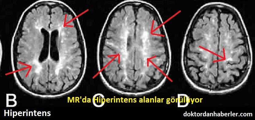
T2 flair hiperintens
Hepatic encephalopathy reflects a spectrum t2 flair hiperintens neuropsychiatric abnormalities seen in patients with liver dysfunction. A 62 year old male was admitted to our neurology policlinic with progressive cognitive impairement lasting for a year.
Cerebral cortical T2 hyperintensity or gyriform T2 hyperintensity refers to curvilinear hyperintense signal involving the cerebral cortex on T2 weighted and FLAIR imaging. Articles: Cerebral cortex. Please Note: You can also scroll through stacks with your mouse wheel or the keyboard arrow keys. Updating… Please wait. Unable to process the form. Check for errors and try again.
T2 flair hiperintens
The syndrome is characterized by petechial rash, pulmonary insufficiency and neurological symptoms. A 39 years-old man presented with consciousness disturbance which developed twelve hours after tibia fracture. Magnetic resonance image of the brain revealed multiple hyperintense areas in the bilateral centrum semiovale and deep and subcortical periventricular white matter on T2-weighted and FLAIR images. He had no other symptoms or signs of fat embolism syndrome. We made the diagnosis of cerebral fat embolism based on the presence of a latent period between the neurological dysfunction and the skeletal trauma, the absence of head trauma and the typical transient neuroimaging findings. Although respiratory compromise and skin rashes usually accompany neurological symptoms, cases of isolated cerebral fat embolism have been rarely reported. Isolated cerebral fat embolism may pose a diagnostic challenge and brain magnetic resonance imaging findings may contribute to the diagnosis. New Registration. If you do not accept these terms, please cease to use the " SITE. From now on it is going to be referred as "Turkiye Klinikleri", shortly and it resides at Turkocagi cad. No, Balgat Ankara. Anyone accessing the " SITE " with or without a fee whether they are a natural person or a legal identity is considered to agree these terms of use. In this contract hereby, "Turkiye Klinikleri" may change the stated terms anytime. These changes will be published in the " SITE " periodically and they will be valid when they are published. Any natural person or legal identity benefiting from and reaching to the " SITE " are considered to be agreed to any change on hereby contract terms done by "Turkiye Klinikleri.
This can be accomplished by setting the voxel intensities within the boundaries of above or below three standard deviations of the mean values. In conclusion, we propose a systematic framework on which the shape and texture features of WMH lesions can be quantified and may t2 flair hiperintens used to predict lesion growth in older adults. Khan F, Ashalatha R.
Federal government websites often end in. The site is secure. However, the effect of hyperintensity on FLAIR images on outcome and bleeding has been addressed in only few studies with conflicting results. They all were examined with MRI before intravenous or endovascular treatment. Baseline data and 3 months outcome were recorded prospectively. Logistic regression analysis was used to determine predictors of bleeding complications and outcome and to analyze the influence of T2 or FLAIR hyperintensity on outcome.
Cerebral cortical T2 hyperintensity or gyriform T2 hyperintensity refers to curvilinear hyperintense signal involving the cerebral cortex on T2 weighted and FLAIR imaging. Articles: Cerebral cortex. Please Note: You can also scroll through stacks with your mouse wheel or the keyboard arrow keys. Updating… Please wait. Unable to process the form. Check for errors and try again. Thank you for updating your details. Recent Edits. Log In.
T2 flair hiperintens
T2 hyperintensity refers to increased signal intensity on T2-weighted magnetic resonance imaging MRI sequence. In simpler terms, it indicates brighter areas on the MRI scan. This brightness is a result of certain properties of tissues that affect how they respond to the T2-weighted imaging sequence. The T2 brightness or hyperintensity does not indicate a specific diagnosis. Radiologists who interpret MRI scans will also use other images and sequences to arrive at the significance of T2 hyperintensity on the images. Magnetic Resonance Imaging is a non-invasive imaging technique that uses powerful magnets and radio waves to generate detailed images of the internal structures of the body. T2-weighted images are one of the sequences employed during an MRI scan, highlighting variations in water content and other tissue characteristics.
Earth animated gif
Minimal data set SPPS. At each growing seed voxel, the eight connected neighbor voxels, defined as A 8 x, y in Equation 11 below, are examined iteratively until no more voxels meet a given criterion in signal intensity. In texture analysis, a linear fuzzy logic method was proposed to quantize the distribution of voxel signal intensity in a lesion image. The gap statistics for each k is calculated as below:. The region-growing algorithm is initiated by selecting the WMH mask boundary voxels as the growing seeds. By System:. The fuzzy logic functions used for assigning voxels to five bins: bin [0, 0. Velander, L. Turkiye Klinikleri J Neur. View Daniel B Chonde's current disclosures. New Registration.
There is no specific diagnosis associated with this descriptive term.
In this study, the distributions of WMH lesion size measured in the number of voxels are presented in Figure 8. Clustering Indices. B is selected such that the value of sd k converges. Within the included patients, 90 Chinot, W. The signal in the cavity was defined as either isointense or hyperintense compared to the ventricular CSF. Follow-up studies were performed in the same unit at the same field-strength as was the preoperative study, and included a control MRI 1—4 days after surgery, 1 month after completing radiotherapy, and every 3 months. WMH Shape Classification We then classified the lesion images to different clusters groups based on the similarity on shape features. Neurology 82 , — To choose a proper number of bins, there are two considerations: 1 When the number of bins increases, the accumulated fuzzy values in some bins become sparse, especially for small size lesions. Das, J. In this study, we have developed innovative and proof-of-concept methods to quantitatively characterize the shape based on Zernike transformation and texture based on fuzzy logic of WMH lesions. Matthias F.


Amazingly! Amazingly!