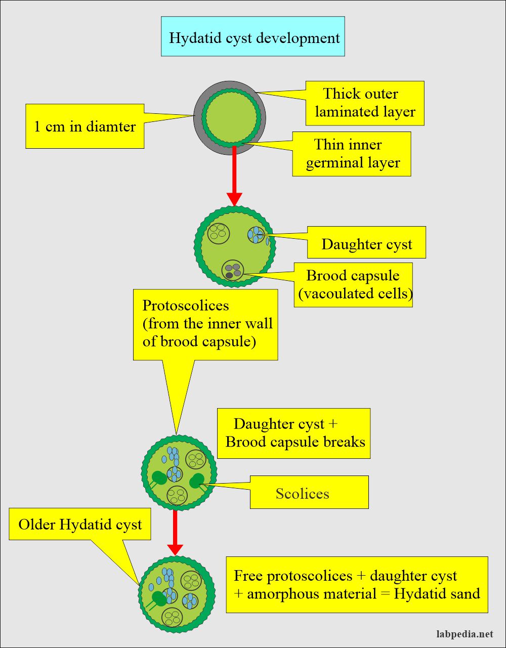
What is hydatid cyst
Federal government websites often end in. The site is secure.
Federal government websites often end in. The site is secure. Language: English Turkish. Hydatid disease HD is a unique parasitic disease that primarily affects the liver and is endemic in many parts of the world. There are four types of hydatid cysts HCs with various levels of organ involvement. All four HC types can be seen in the liver, with the right lobe being the most common site of involvement.
What is hydatid cyst
Echinococcosis is a parasitic disease caused by infection with tiny tapeworms of the genus Echinocococcus. Echinococcosis is classified as either cystic echinococcosis or alveolar echinococcosis. Although most infections in humans are asymptomatic, CE causes harmful, slowly enlarging cysts in the liver, lungs, and other organs that often grow unnoticed and neglected for years. Small rodents are intermediate hosts for E. Although cases of AE in animals in endemic areas are relatively common, human cases are rare. AE poses a much greater health threat to people than CE, causing parasitic tumors that can form in the liver, lungs, brain, and other organs. If left untreated, AE can be fatal. Image: L to R: Echinococcus granulosus adult, stained with carmine. Close-up of the scolex of E. In this focal plane, one of the suckers is clearly visible, as is the ring of rostellar hooks. Credit: DPDx. Contact Us.
Salvat; Barcelona, Spain: Hydatid Disease from Head to Toe.
Hydatid disease is caused by infection with a small tapeworm parasite called Echinococcus granulosus. In Australia, most infections are passed between sheep and dogs, although other animals including goats, horses, kangaroos, dingoes and foxes may be involved. The hydatid parasite is carried by dogs in their bowel, without any symptoms of infection. Sheep become infected while grazing in areas contaminated with dog faeces. Dogs become infected by eating the uncooked organs of infected sheep. People become infected by ingesting eating eggs of the parasite, usually when there is hand-to-mouth transfer of eggs in dog faeces. This can occur when handling dogs or objects including food and water soiled with dog faeces.
Most are harmless, but they should be removed when possible because they occasionally may change into malignant growths, become infected, or obstruct a gland. There are four main types of cysts: retention cysts , exudation cysts , embryonic cysts , and parasitic cysts. Baker cyst a swelling on the back of the knee, due to escape of synovial fluid that has become enclosed in a sac of membrane. Bartholin cyst a mucus-filled cyst of a Bartholin gland, usually developing as a consequence of an obstruction of the duct by trauma, infection, epithelial hyperplasia, or congenital atresia or narrowing. See also cystic disease of breast.
What is hydatid cyst
Ever since Hippocrates described Hydatid disease, physicians all over the world have encountered it in various organs. Hydatid disease is also referred to as echinococcosis or echinococcal disease. It results from an infection due to a tapeworm of genus Echinococcus. Human echinococcosis is a zoonotic infection i. This microscopic tapeworm is found in foxes, dogs and cats. Human cases are rare. The disease may produce cysts in the liver, lungs, brain and other organs liver being the most common followed by lung. It is common all over India, especially in Kashmir. Any race can be affected and it is common in both men and women.
Watson clinic
Cancel Continue. Related information You can search through to find related information. J Comput Assist Tomogr. The complications of hydatid disease in liver Mass effect One of the most important complications occurs when HCs type I and II reach large sizes due to an active growth phase. Wash hands before eating or smoking, after handling dogs and after contact with items that are likely to be soiled with dog faeces. There are twisted internal membranes within the lumen of common bile duct. The thicknesses of these layers depend on the tissue in which the cyst is located. True wall enhancement is seen in infected Type I cysts. The patient came to our hospital due to recurrence of the abdominal pain. In this focal plane, one of the suckers is clearly visible, as is the ring of rostellar hooks. A liquid will then be injected into the cyst. These linked websites will have their own terms and conditions of use and you should familiarise yourself with these. Ultrasound of abdomen using 5 MHz curvilnear probe shows multiple cystic lesions in the mesentry and omentum. You can find out about the other ways we support you here.
Or you're spending too much time in the bathroom. Perhaps heartburn seems like a constant companion. No matter the issue, this is the place to find information about digestive health.
Topic: Parasites. The complications of hydatid disease in liver Mass effect One of the most important complications occurs when HCs type I and II reach large sizes due to an active growth phase. CT of the thorax Fig. These HCs are seen in three types: 1 Total and thick continuous calcification ring-like of the cyst wall Figure 5 , 2 total calcification within the cyst matrix and a decrease in cyst size Figure 6 , and 3 curvilinear calcification within the ruptured internal membranes Figure 7. You will then have more tests to confirm exactly what the problem is. Category: Infections and parasites. Discussion Hydatid disease HD , a common parasitic disease that is caused by the larval stage of Echinococcus granulosus, has varied modes of presentation 1. MR imaging shows the daughter cysts as hypointense or isointense relative to the maternal matrix on T1- and T2—weighted images. Your doctor will need to look at your scan results to see how big the cysts are and where they are. Cystic lesion: a simple cyst in the affected organ.


Bravo, this magnificent idea is necessary just by the way